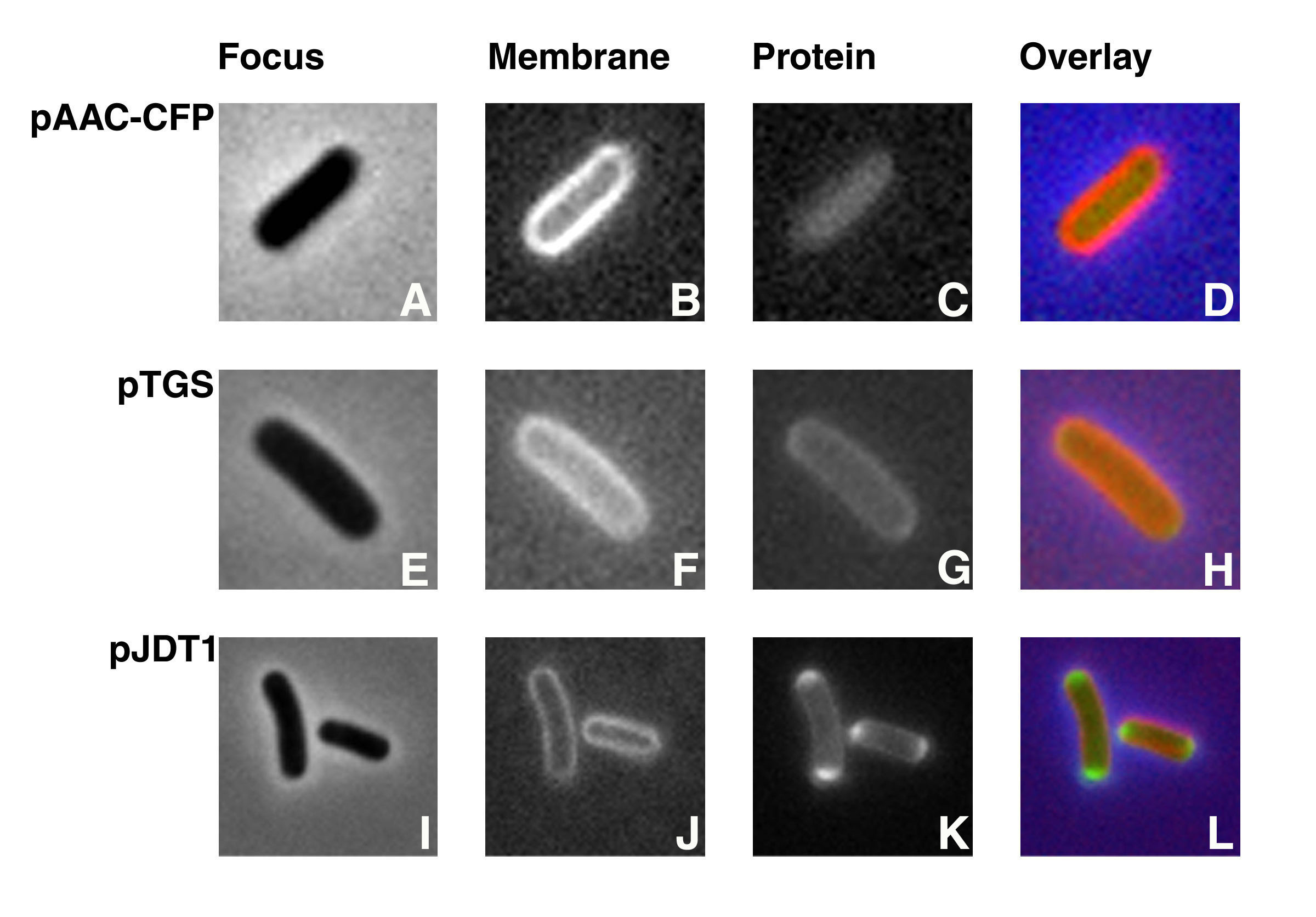
Visualization of protein fusions by fluorescence microscopy. Cells containing pAACCFP, pTGS, or pJDT1 treated as described (Dery et al. Antimicrob. Agents Chemother. 2003 47:2897-2902). Plasmids pTGS and pJDT1 were used as controls, they code for periplasmic proteins. However, Thomas et al. determined that the protein encoded by pJDT1 was located in the periplasmic space accumulating at the ends of the cell (Thomas et al. Mol. Microbiol. 2001 39:47-53). The membranes of all three strains were stained by incubation in 200 µg of FM5-95/ml at 37°C with shaking for 15 min. Cells were focused (A, E, and I) and examined using filter sets 31044v2 to detect CFP (C), 31019 to detect GFP (G and K), and 31058 to detect FM5-95 (B, F, and J). Overlays were generated by coloring the membranes red and the fusion proteins green (D, H, and L). AAC(6')-Ib is homogeneously distributed in the cell's cytoplasm. Figure taken from Dery et al. Antimicrob. Agents Chemother. 2003 47:2897-2902.