source of image: Eugene Weber, California Academy of Sciences
Notes for Chapter 12:
Porifera ("Mesozoa and Parazoa")
Click link to return to Lecture
Schedule
or back to Chapter 11
or ahead to Chapter 13
Chapter 12 Assignment:
Ch. 12: 240-241, 243-252; RQ-12: 3-5, 8-11
(Porifera only, not Mesozoa, Placozoa)
Introduction: The Advent of Multicellularity
source
of image: Eugene Weber, California Academy of Sciences
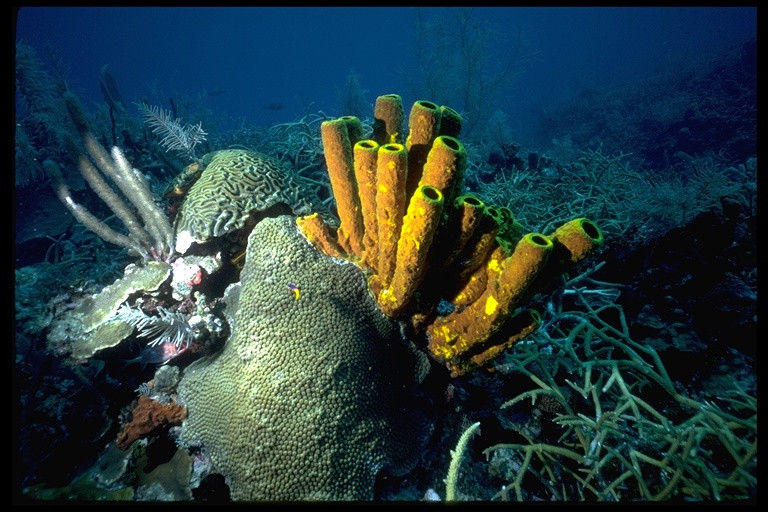
Featured Organism:
Aplysina fistularis
Links:
1
- 2
- 3 - 4;
Other Aplysina spp.: 1
- 2
- 3
- 4
I. Origin of Metazoa
This section is badly out of date. To gain
a better view of traditional vs. current estimates of metazoan phylogeny, I
recommend that you instead view these
diagrams for a comparison of older
views of animal relationships with a more up-to-date tree
of animal relationships based on DNA sequence. For
an overview of the early fossil record of animals through time
click here,
or see this
article for a more detailed overview
including recent fossil and developmental discoveries.
Key Terms: syncytical ciliate hypothesis (abandoned), colonial flagellate hypothesis (leading hypothesis, especially with something like a choanoflagellate colony giving rise to the first metazoan), polyphyletic origin (no evidence, instead, there is much evidence that Metazoa is monophyletic), cellular level of organization, incipient tissues
Metazoan synapomorphies (shared-derived novelties):
Multicellularity (but other eukaryotes also multicellular)
Extracellular matrix (ECM) that provides structural support
Intercellular cytoplasmic communication
Septate and desmosomal junctions between cells
Extracellular production of cross-linked collagen
The following junction/tissue type details should not be memorized:
ECM are made of gelatinous matrix - glycosaminoglycans
(GAGs)
GAGs are infused with fibers - especially collagen, but also others
Examples of junction types:
occluding - seal passage of material across cell sheets
(include septate and tight junctions)
adhesion - including spot and belt desmosomes
communicating - permit passage of material through tubes
(as in gap juctions)
or through electrical/molecular system (as in
neurotransmitter-synapse junctions)
Sponges do not have communicating junctions
Two general categories of metazoan tissues:
Connective
- cells not in sheets
- not sealed by occluding junctions
- rich in ECM
Epithelial
- sheets of cells
- polarized into apical and basal portions
- sealed (usually) by occluding junctions
- nervous tissues - highly modified type
Phylogeny (simplified):
Metazoa
Porifera (Sponges)
Eumetazoa (tissues, expanded gut)
Cnidaria (radial symmetry)
Bilateria (bilateral symmetry)
II. Mesozoa (skip, some of these were once thought to resemble a possible metazoan ancestor but recent molecular studies instead provide evidence that at least some are secondarily simplified triploblastic metazoans)
III. Placozoa (skip, these simple animals known mostly from the glass walls of tropical aquaria are apparently metazoans whose affinities may lie with eumetazoans, cnidarians, or bilaterians, so they do not appear to be "ancestors" of other metazoans)
IV. Porifera
(sponges)
More
Links
A. Sponge diversity (about 5,000 species)
Silicea (siliceous "glass" spicules)
Hexactinellida ("glass sponges," mostly deepwater)
Demospongia (common, diverse)
Calcarea (calcareous spicules)Recent molecular studies have surprisingly found evidence for a more basal position of Silicea within metazoans. By this view, Calcarea are sister taxon of Eumetazoa (all metazoans except for sponges), so "Porifera" is paraphyletic and must be abandoned. If this phylogenetic hypothesis is accurate, it strengthens the notion that the ancestor of all metazoans was sponge-like. It also suggests that typical textbook emphasis on a grade of increasing complexity within sponges (e.g., Fig. 12-5), from asconoid, to syconoid, to leuconoid, has nothing to do with the origin of eumetazoans from sponge-like ancestors. This is because our closest relatives, calcareans, generally have the more simple asconoid body type.
B. How a sponge works
Aquatic animals
Can be soft-bodied but most firm to woody
– internal skelton of many tiny calcareous or siliceous spicules
– many also have tough sponge fibers
Sponges are perforated by a nework of canals
– water moves to chambers
– moves to chambers lined with "collar" cells (Choanocytes)
– exits through a corridor to one or more large oscula
Sponges are highly specialized to filter feed.
– One sponge can filter multiple liters of water
Sponges have tissues
– Complex array of cell types
– No organs or nervous system
–Only two "systems"
canals/chambers
skeleton
See Fig. 12-10: Note how the choanocyte cell (collar cell) traps food particles through the action of a beating flagellum, drawing water through collar microvilli. Note that incurrent flow is through tiny ostia pores, whereas relatively fewer collar cells share exhalent oscula pores. The sponge body is more or less complicated (Fig. 12-5). Note that there are loosely organized "tissue layers" in a sponge, with different types of cells, including archaeocytes, pinacocytes, and collencytes, all suspended in a gelatinous mesohyl matrix, typically with a skeletal framework of spicules, and usually also spongin fibers (Fig. 12-9). (Bath sponges fortunately have spongin but no spicules.)
C. What did sponges evolve from?
Note that sponges feed by cellular digestion. They have no gut. A gut is a synapomorphy of Eumetazoa, the clade of all metazoans besides sponges. In this regard sponges are primitive. (In other aspects they are highly specialized.) Sponge choanocyte cells very closely resemble the "protist" organisms we covered in Chapter 11 called choanoflagellates (e.g., Codosiga, Fig. 11-14). For descriptions of some other choanoflagellates, see Monosiga or Salpingoeca. Despite their resemblance to choanoflagellates, sponges are more closely related to us (and other eumetazoans) than they are to choanoflagellates. The evidence for this statement is the synapomorphies we share with sponges, lacking in choanoflagellates. These derived similarities are most parsimoniously explained by the hypothesis that we share a common ancestor with sponges that is more recent than the one either of us shares with choanoflagellates.
Click link to return to Lecture
Schedule
or back to Chapter 11
or ahead to Chapter 13
This page created 9/16/01 © D.J. Eernisse, Last Modified 9/16/01, Links Last Completely Checked 9/16/01